


PSMA-PET imaging for prostate cancer: Exploring gallium-68 in clinical practice
Telix spoke with a panel of global experts to examine the rapid evolution of gallium-68 (68Ga) PSMA-PET imaging in prostate cancer and to ask where the technology goes next.
The experts said that although PSMA-PET imaging is now established as the standard of care for identifying and staging prostate cancer, continued innovation with 68Ga is creating new opportunities to improve patient care.
The evidence base
A decade that changed everything
PSMA-PET imaging for prostate cancer (positron emission tomography targeting prostate-specific membrane antigen) is now used across several stages of care, including staging, biochemical recurrence, and selection for radioligand therapy, with use in initial diagnosis under active investigation. In under a decade, PSMA-PET has moved from a research tool used in a handful of centers to a widely adopted standard in prostate cancer staging and management.
To understand where PSMA-PET imaging goes next, Telix spoke with Rodney Hicks, MD; Paul Yonover, MD; Telix Group Chief Operating Officer Dr. Darren Patti; and Telix Group Chief Medical Officer David N. Cade, MBBS
 Rodney Hicks, MD
Chief Medical Officer and Nuclear Medicine specialist, Melbourne Theranostics Innovation Centre
Rodney Hicks, MD
Chief Medical Officer and Nuclear Medicine specialist, Melbourne Theranostics Innovation Centre
 Paul Yonover, MD
Chief Data Officer and Director of Clinical Navigation, UroPartners, Chicago
Paul Yonover, MD
Chief Data Officer and Director of Clinical Navigation, UroPartners, Chicago
 David N. Cade, MBBS
Telix Group Chief Medical Officer
David N. Cade, MBBS
Telix Group Chief Medical Officer
 Dr. Darren Patti
Telix Group Chief Operating Officer
Dr. Darren Patti
Telix Group Chief Operating Officer
Professor Hicks and Dr. Yonover are independent expert speakers and not employees of Telix. Views are the speakers’ own.
AccuracyHow accurate is PSMA-PET for prostate cancer?
Professor Hicks installed one of the first PET-CT cameras in Australia at the Peter MacCallum Cancer Centre in 2001 and has practiced PSMA-targeted theranostics for over a decade.
Across a review of 76 published articles in more than 5,000 patients, gallium-68 (68Ga) PSMA-11 PET has demonstrated overall sensitivity of at least 80% and specificity of at least 90%1. The technology shows high true positive detection rates for regional and distant metastases, including in bone and soft tissue, and high diagnostic performance for micrometastatic disease as small as 2 mm and at PSA values as low as 0.02 ng/mL2-5. Studies have consistently shown high reproducibility and inter-reader agreement6,7.
Gallium-68 PSMA-11 PET can inform care across these stages: disease staging, biochemical recurrence after definitive treatment, and selection for radioligand therapy, with its role in initial diagnosis under active investigation (fig. 1). More recently, gallium-based PSMA imaging has begun to be used in the assessment of therapeutic response.

Recent data adds to this. Results from Telix’s Phase 3 registration study of 68Ga PSMA-11 PET in a Chinese patient population, first announced in December 2025, demonstrated an overall patient-level positive predictive value (PPV) of 94.8% (95% CI: 85.9%–98.2%) for tumor detection, well above the predefined threshold of 60%8. Region-level PPV was 100% in the prostate bed, extra-pelvic soft tissue, lymph nodes and organ metastases, and 87% in bone metastases. More than two-thirds of patients (67.2%) had a change in their treatment plan following PSMA-PET imaging compared with the initial plan at baseline, an indicator of its potential influence on clinical management8.
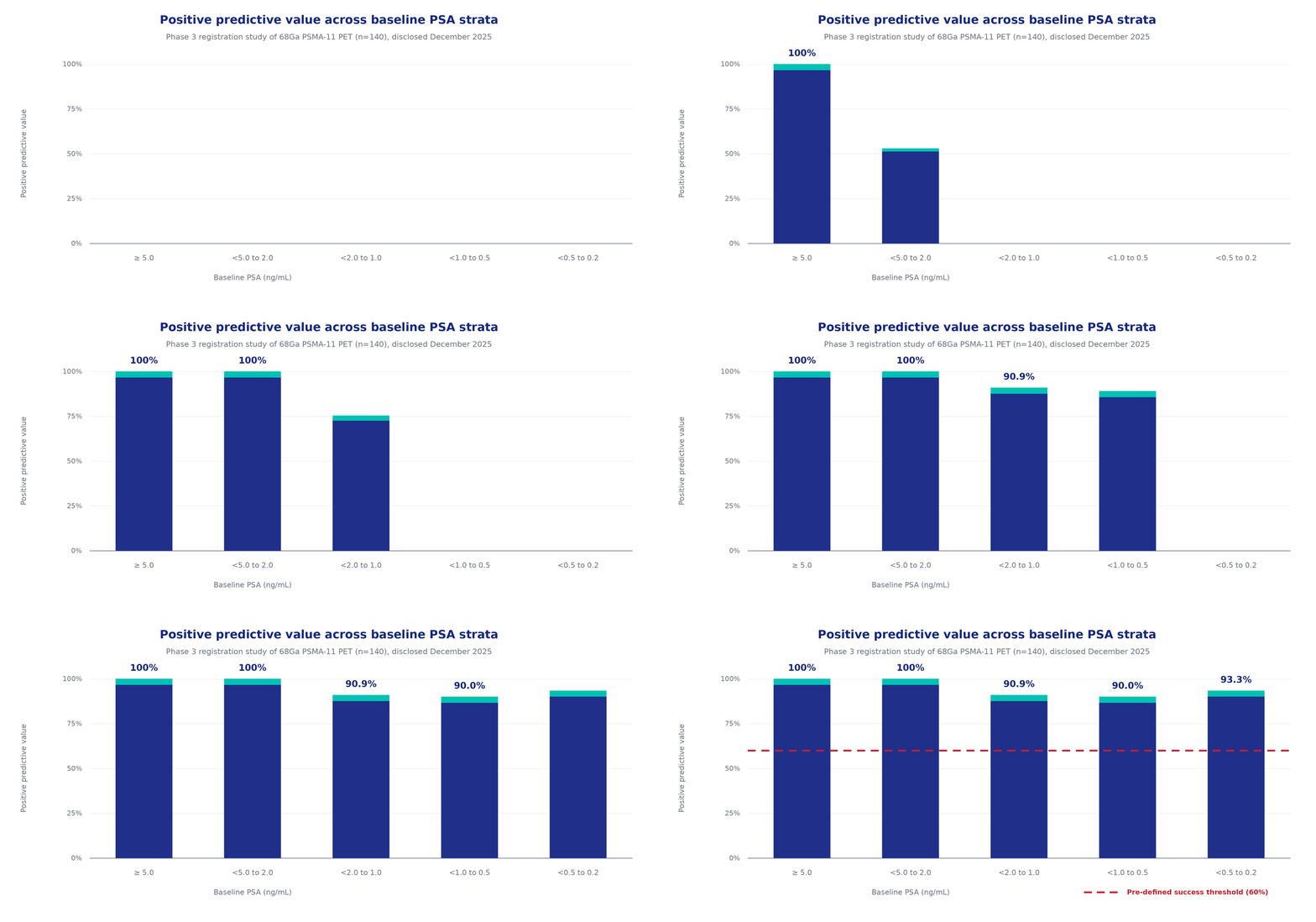
“The area where we’re seeing the greatest impact is actually in those very low PSA levels where traditionally people haven’t been doing it [PSMA-PET],” Professor Hicks observed. In biochemical recurrence, finding disease at these sub-0.2 ng/mL PSA values matters increasingly for selecting patients who may benefit from salvage locoregional therapies, and for sparing those whose disease turns out to be more widespread than conventional imaging suggests.
TechniqueAdvanced methods for improving PSMA-PET detection
For Professor Hicks, optimizing diagnostic performance comes down to a simple framework. A lesion can be missed on PSMA-PET for three reasons, and each has its own solution.
A question of technology and technique: extended field-of-view digital PET cameras and optimized acquisition.
Lesions near the bladder or major vessels, addressed with late imaging and dynamic diuretic protocols.
Low PSMA expression on the tumor itself, which calls for alternative tracers or targets altogether.
The first two are mostly questions of technology and technique. Professor Hicks pointed to extended field-of-view (EFOV) digital PET cameras such as the Siemens Biograph Vision Quadra, which he has installed at the Melbourne Theranostics Innovation Centre, against the conventional scanners that dominated the field until recently. “The Quadra, having a field of view of over a metre and very high time-of-flight resolution and very sensitive digital detectors, is about ten-fold higher [in sensitivity] than the best [conventional] digital camera that Siemens themselves make, which itself is about twice as sensitive as most non-digital time-of-flight cameras, which are themselves twice as sensitive as analogue cameras without time of flight.” Taken together, that is a sensitivity advantage of somewhere between ten and forty times over older equipment.
A patient had been referred for salvage pelvic radiotherapy on the basis of an elevated PSA of around 0.4 ng/mL, with a conventional scan suggesting possible locoregional disease. On the Quadra, using an optimized late post-diuretic imaging protocol, the picture was completely different (Fig. 3). Disease was widespread but the prostate bed was clear, and the patient would have gained nothing from pelvic radiotherapy. “Huge impact on patients,” he said.
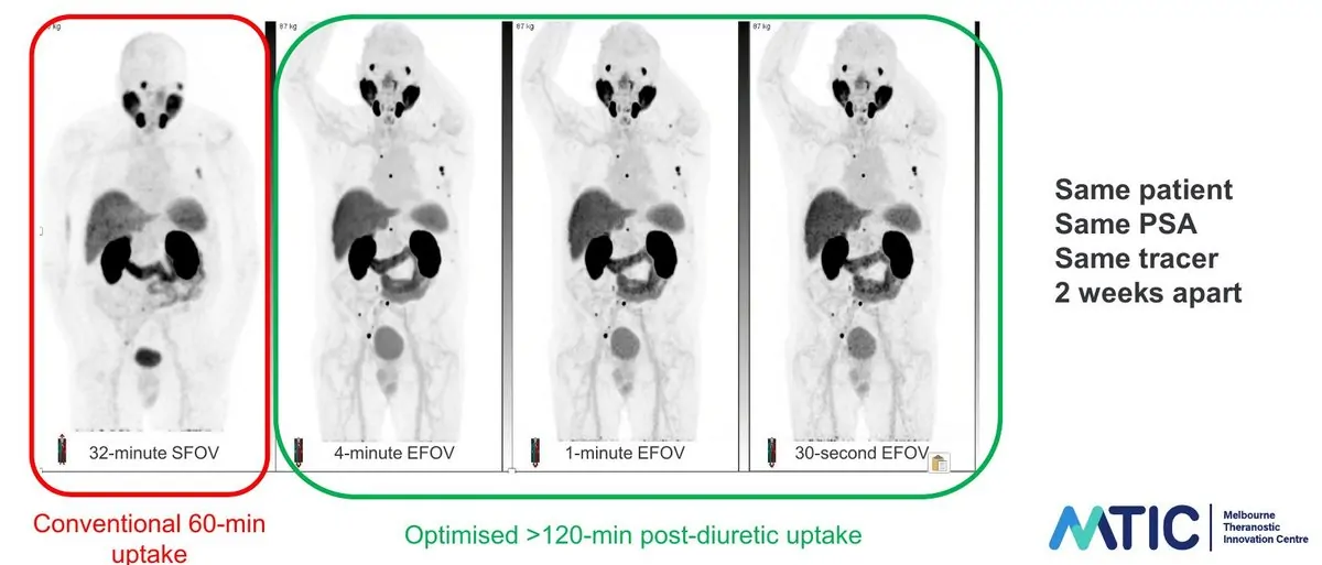
Technique matters as much as the hardware. Imaging later, beyond 120 minutes post-injection and with dynamic diuretic protocols to clear bladder activity, substantially improves detection of lesions sitting close to the bladder and pelvic vessels. Artificial intelligence-assisted image processing, Professor Hicks argued, will deliver a further jump in sensitivity for small-volume disease.
The third barrier, too little target, calls for a different approach altogether. PSMA expression is not uniform across the disease spectrum. At the high end of Gleason grading, particularly in pattern 5 disease, PSMA expression can be absent. “Those are genuine card-carrying cancers that use glucose,” Professor Hicks noted, and FDG-PET has shown real utility in this subgroup. In lower-grade disease around the Gleason 6-to-7 transition, alternative targets such as the gastrin-releasing peptide receptor (GRP-R) are overexpressed and represent an active area of tracer development with both diagnostic and therapeutic potential. These are limitations of the PSMA target itself rather than of 68Ga as an isotope, a distinction that bears on how the field develops.
Clinical practicePSMA-PET in high-volume urology practice
As a practising urologist, Dr Paul Yonover brought the perspective of high-volume clinical practice, where most patients encounter this technology.
UroPartners is a key affiliate within a large, nationally distributed U.S. private urology practice that Dr. Yonover describes as a “closed ecosystem”, covering screening, diagnosis, pathology, in-office dispensing, linear accelerators, brachytherapy and theranostics within a single organization. With multiple PET-CT scanners running across major U.S. cities and more planned, it performs thousands of PSMA-PET scans a year.
Same-day scanning is non-negotiable. Prior authorization, patient transport, no-show rates, scan scheduling and data turnaround are all points of friction in a system handling enormous throughput. Choosing an imaging platform, including the isotope, tracer, camera and protocol, is therefore an operational and economic decision as much as a clinical one.
On that basis the practice has committed to 68Ga. “We’ve already executed a business plan around gallium scanning,” Dr. Yonover said, and “the bar has been set extremely high by 68Ga and there would have to be an extremely compelling reason to change course in the future.” The depth of the gallium evidence base, built over five to ten years of high-volume clinical use, underpins that commitment as much as any logistical factor. “As a clinician and a business owner, I’m much more interested in novel targets” than novel isotopes, he added, pointing to the known limitations of PSMA targeting in certain disease phenotypes as a more pressing unmet need than swapping isotopes.
The clinical impact on his own practice has been considerable. “It’s hard to overstate the impact [molecular imaging] has had, just in the last four or five years when it’s really come to the fore here in the United States and how it’s been disruptive in a good way,” he said. Being able to go into surgery with real confidence about disease extent, and to avoid the surprise of finding occult nodal involvement or extracapsular disease, has changed how he plans operations. “When you’re getting closer to truth, you can actually just make better decisions.”
Supply chainProven at scale
Every PSMA-PET scan depends on a supply chain that can deliver a radioactive tracer to thousands of patients reliably, across imaging centres that may be hundreds of miles apart, a logistical problem the wider healthcare system has sometimes underestimated.
Dr. Darren Patti, Telix’s Group Chief Operating Officer, knows this side of the business well, having held senior roles at Sofie and in PET tracer manufacturing and radiopharmacy network operations across North America. He is clear about the strength the gallium supply chain now has.
“Now that we have multiple commercial products on the market for a number of years, this has really given opportunity for the gallium isotope supply chain to mature.” Several generator manufacturers now provide consistent access, and the supply chain from germanium-68 feedstock through to finished drug product has become more vertically integrated, reducing the risk of interruption. A generator on site or nearby gives clinics on-demand availability and the same-day scheduling flexibility high-volume practices depend on.
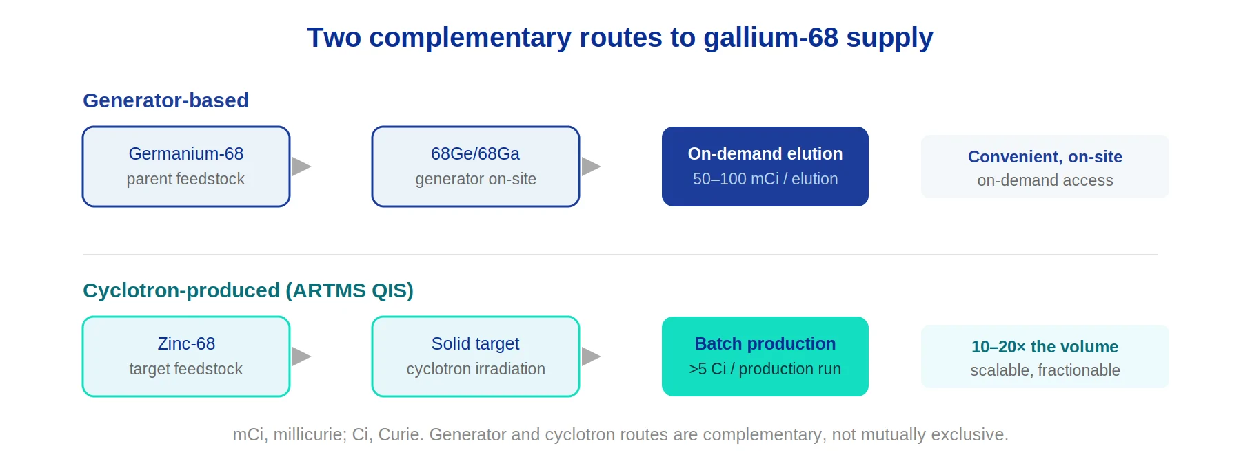
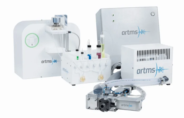
Generator-based production is now being augmented, and where needed substantially scaled, by cyclotron-produced gallium. Telix’s ARTMS platform, using the QUANTM Irradiation System (QIS), achieves production yields above 5 curie per run from solid target cyclotron systems. By comparison, conventional generators typically produce 50 to 100 millicurie per elution, so cyclotron production can deliver ten to twenty times the isotope volume. “That really opens up a lot of flexibility in terms of how radiopharmacies and those producing finished drug product can address the market,” Patti said. Fractionating cyclotron-produced gallium out to several downstream centres extends geographic reach further still.
With scanning volumes in the prostate cancer imaging market forecast to potentially double by the early 2030s, pairing a proven generator network with scalable cyclotron production is meant to meet that demand, giving what Patti calls “a flexible option that can be tailored to a specific market need.”
The isotope questionGallium-68 vs novel isotopes: what physicians actually want
Both Professor Hicks and Dr. Yonover see a defined role for novel isotopes, but a bounded one.
That role is in specific clinical niches, particularly where a longer-lived radionuclide offers diagnostic advantages, such as late imaging for predictive dosimetry in patients being considered for radioligand therapy where PSMA target intensity is borderline. Imaging at 24 hours or beyond, and deriving a time-activity curve for individual dose estimation, is a scientifically meaningful capability in that setting.
“I see something like copper as a niche product,” Dr. Yonover said. “There are going to be times where I think it’s probably going to be useful in that space, but I’m personally much more interested in different targets than I am in different isotopes.” Professor Hicks added that “in most patients within one to two hours, you’ve got all the information you need for therapeutic pathway planning” with 68Ga PSMA-11.
What physicians want from the future, both agreed, is to build on what already works: optimized acquisition protocols, smarter AI-assisted image processing, better cameras, and above all novel molecular targets that close the known gaps in PSMA expression across the prostate cancer phenotypic spectrum.
What comes nextThe future of PSMA-PET: biopsy guidance, response monitoring and earlier detection
Telix’s Phase 3 BiPASS™ study is evaluating pre-biopsy 68Ga-PSMA-PET combined with multiparametric MRI in men assessed for clinically significant prostate cancer. The hypothesis: improve biopsy yield, reduce unnecessary biopsies in low-risk patients, and provide whole-body staging in a single visit. “One-stop shopping,” in Dr. Yonover’s words.
Professor Hicks stressed how MRI and 68Ga-PSMA-PET complement each other. The two are not competing modalities; they assess different biological dimensions of a heterogeneous disease. Areas where both MRI and biopsy have inherent limitations, such as the anterior fibromuscular stroma and lesions at the very base of the prostate close to the bladder, are exactly where 68Ga-PSMA-PET has identified disease that other methods missed. Biopsy-negative, strongly PSMA-positive cases happen, and they matter clinically.
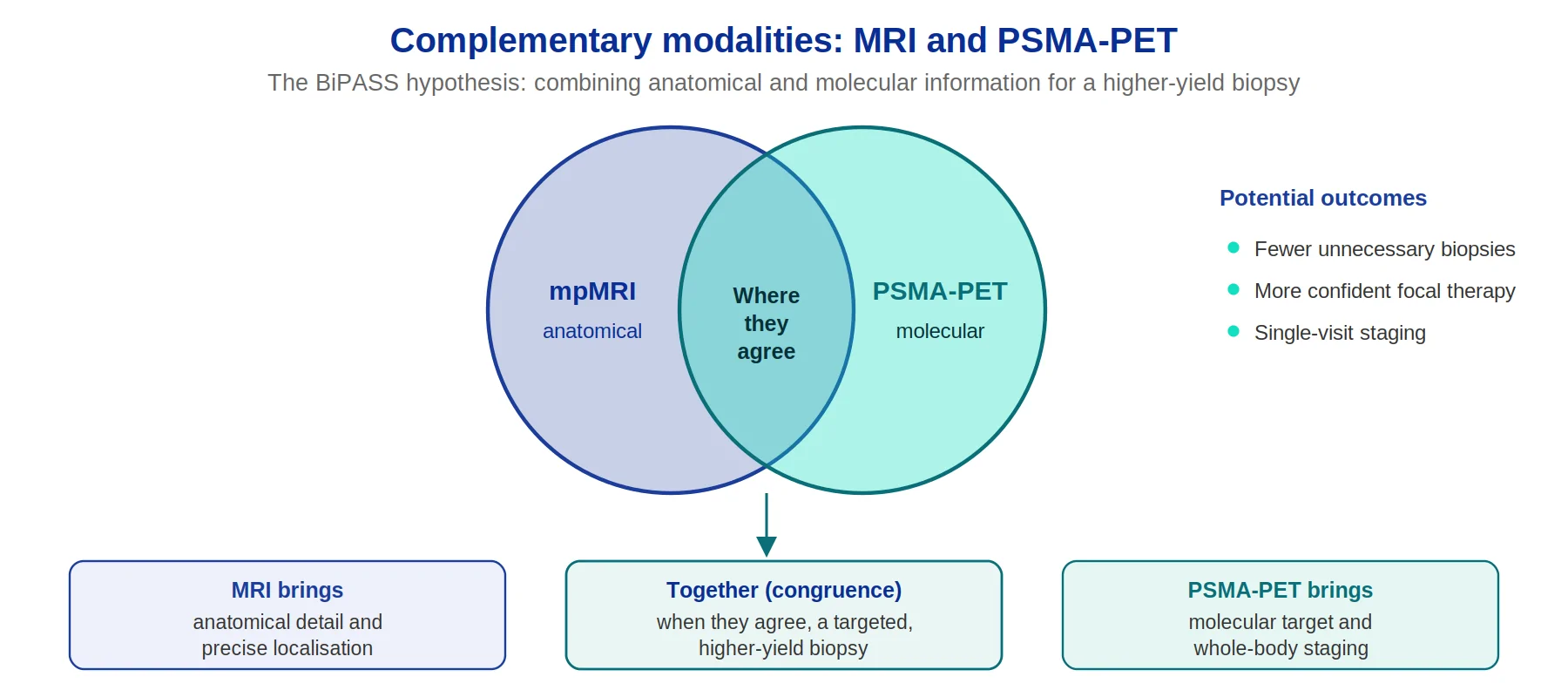
Dr. Yonover, who has performed BiPASS biopsies guided by PSMA-PET in his own practice, sees implications for focal therapy, a modality he believes has been underused partly because of uncertainty about where disease sits. “If I have a higher yield, more accurate biopsy, particularly if there’s congruence between my molecular image and my MRI, I think the field of focal therapy will start to bloom,” he said, “which is going to be an advantage particularly for our younger patients.”
Response monitoring is another frontier. Post-treatment follow-up in prostate cancer still leans heavily on serial PSA measurement and conventional imaging that is poorly suited to detecting small-volume residual or recurrent disease. Bringing 68Ga-PSMA-PET into response assessment, recently recognized in updated Prostate Cancer Working Group guidance, raises the prospect of earlier, more sensitive detection of treatment failure and more rational decisions about escalating or de-escalating therapy. As Professor Hicks put it: “If you can’t see it, you can’t measure it, you can’t monitor it.”
The physicians Telix spoke with regard gallium-68 PSMA-11 imaging as one of the most significant diagnostic advances in prostate cancer of the past decade. The evidence base has grown substantially, the supply infrastructure is maturing and scaling, and clinicians who use the technology daily are applying it in earlier and more nuanced settings. For them, the open question is no longer whether it belongs in the clinic, but how far it can go.
Independent expert disclosure
Professor Rodney Hicks and Dr Paul Yonover took part in this interview as independent expert speakers. They are not employees of Telix Pharmaceuticals, and the views expressed are their own. Any investigational products or applications discussed, including the BiPASS™ trial and expanded indications for PSMA-PET imaging, have not received regulatory approval and are subject to ongoing clinical evaluation.
References
- Purysko AS, et al. Appl Radiol. 2024;53(2):22–35.
- Rauscher I, et al. J Nucl Med. 2020;61(1):51–57.
- Kroenke M, et al. J Nucl Med. 2021;62(8):1082–1088.
- Burgard C, et al. Cancers (Basel). 2023;15(5):1376.
- Hagens MJ, et al. J Nucl Med. 2022;63(10):1531–1536.
- Giesel FL, et al. Eur J Nucl Med Mol Imaging. 2015;42(12):1794–1800.
- van Leeuwen PJ, et al. BJU Int. 2017;119(2):209–215.
- Telix ASX disclosure, December 22, 2025. 68Ga PSMA-11 China Phase 3 Registration Study Clinical Study Report, December 2025.